Articles · Health research
Children are not small adults.
Radiologists with Texas affiliations warn that pediatric imaging is underrepresented in AI research. A separate MRI study illustrates why performance must be tested beyond the original patient group.
Read the story
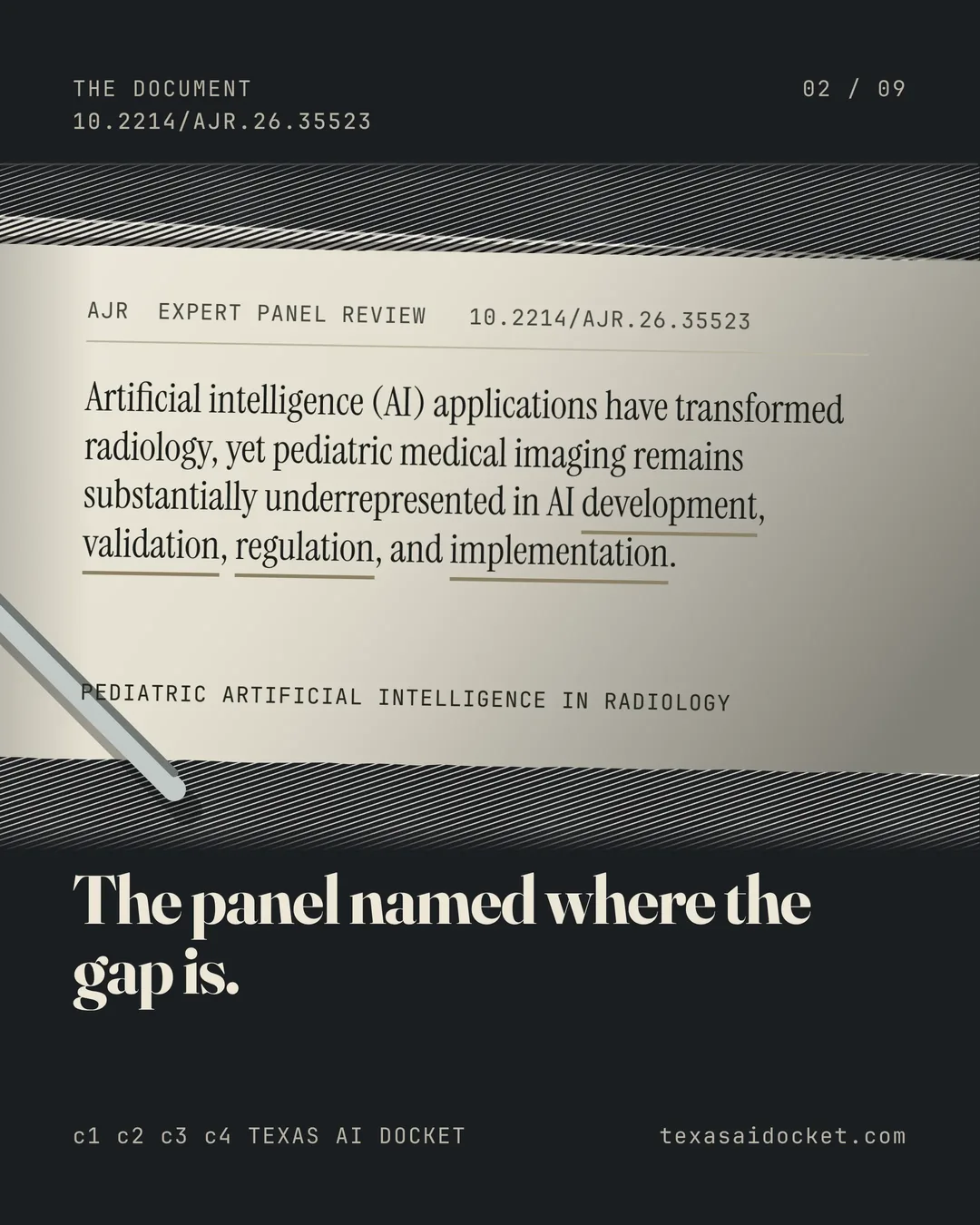


An AI model built around adults does not automatically meet children's needs. An expert panel review argues that pediatric imaging remains underrepresented in AI development, validation, regulation and implementation.
The panel includes radiologists affiliated with Texas Children's Hospital and UT Southwestern. Its document is a narrative review with recommendations, not a new trial measuring a pediatric product's safety or effectiveness.
Children require their own evidence
The review identifies scarce public datasets, fragmented institutional records and insufficient external validation among the obstacles. It also raises concerns about consent, off-label use of adult-trained models and surveillance after deployment.
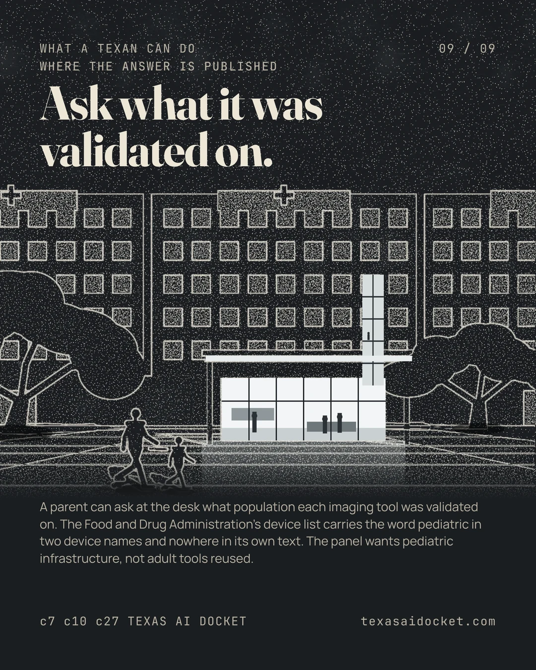
Its proposed response is dedicated pediatric infrastructure and standards. That recommendation identifies work still needed. It is not a finding that every existing imaging tool is unsuitable for every child.
A separate test of generalization
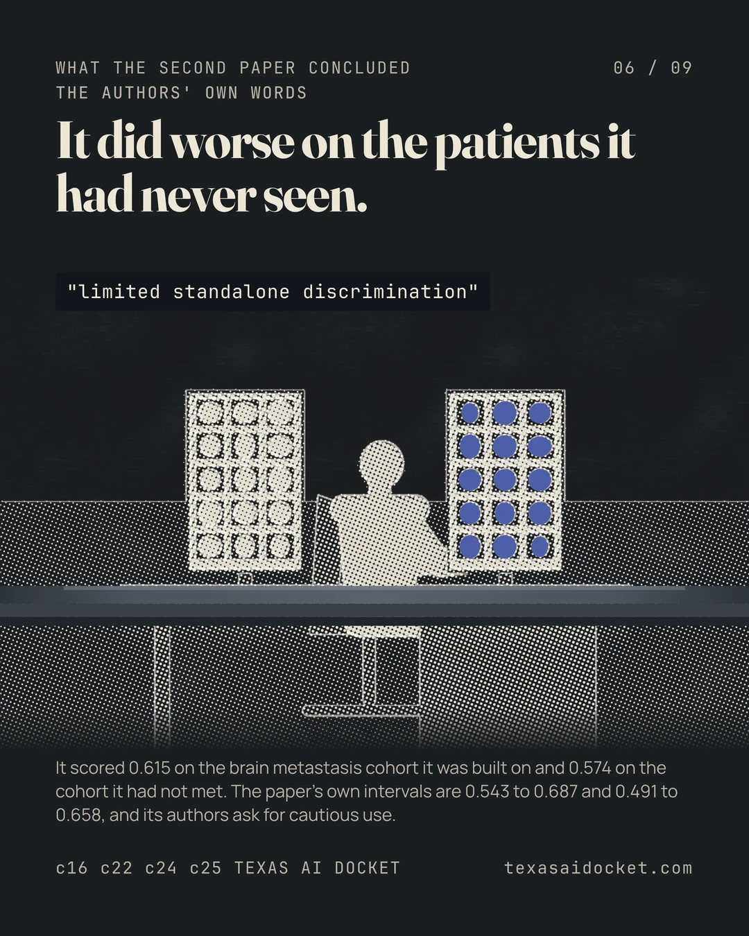
A separate MRI radiomics study developed a survival model in one patient cohort and tested it without refitting in another. Performance was lower in the independent cohort. The authors described limited standalone discrimination and urged cautious use.
That paper is not a pediatric trial. It illustrates a related evidence problem without resolving the review's child-specific questions. Stronger results in the development group do not guarantee the same performance elsewhere.
What remains to be demonstrated
Evidence needs to match the intended patient population and use. The review's priorities and the separate study's limitations both point toward validation, not an assumption that a successful model travels unchanged between settings.
Sources for this story
- PubMed record 42714442, AJR. American journal of roentgenology
- PubMed record 42716711, AJNR. American journal of neuroradiology
- Artificial Intelligence-Enabled Medical Devices, U.S. Food and Drug Administration
Claim-by-claim verification · 27 claims
The review's title names it an expert panel review of pediatric artificial intelligence in radiology.
Pediatric Artificial Intelligence in Radiology: Unmet Needs and Next Steps-AJR Expert Panel Review.
The review carries an electronic article date of September 9th, 2026.
<ArticleDate DateType="Electronic"><Year>2026</Year><Month>09</Month><Day>09</Day></ArticleDate>
The review is registered under its own digital object identifier.
<ELocationID EIdType="doi" ValidYN="Y">10.2214/AJR.26.35523</ELocationID>
The review opens by stating that pediatric medical imaging remains substantially underrepresented across AI development, validation, regulation and implementation.
Artificial intelligence (AI) applications have transformed radiology, yet pediatric medical imaging remains substantially underrepresented in AI development, validation, regulation, and implementation.
The panel states that children change continuously as they grow, so a model fitted to adults is not the model a child needs.
Unlike adults, children go through continuous physiologic and anatomic changes that require age-specific models trained on representative developmental data.
The panel names five things that limit pediatric AI.
However, pediatric AI is limited by scarce publicly available datasets, fragmented institutional data, rare diseases, heterogeneous reporting practices, and insufficient external validation.
The panel names off-label use of adult-trained AI models on children as an ethical and regulatory concern.
Ethical and regulatory challenges are also a concern in children, including consent for secondary data use, off-label use of adult-trained AI models, and the need for postdeployment surveillance.
The panel states that reimbursement is misaligned and must be optimized before innovation can follow.
Additionally, reimbursement is misaligned and must be optimized to allow innovation.
The document describes itself as a narrative review proposing practical priorities rather than as original research.
This AJR Expert Panel Narrative Review examines the current landscape of pediatric AI in radiology and proposes practical priorities to support its safe and equitable adoption.
The panel's central recommendation is an implementation roadmap for a dedicated pediatric AI infrastructure and standards.
The panel gives key recommendations, emphasizing the importance of an implementation roadmap to establish a dedicated pediatric AI infrastructure and standards that are essential to ensure diagnostic accuracy, workflow efficiency, and optimal clinical outcomes for children while minimizing bias and protecting patient safety.
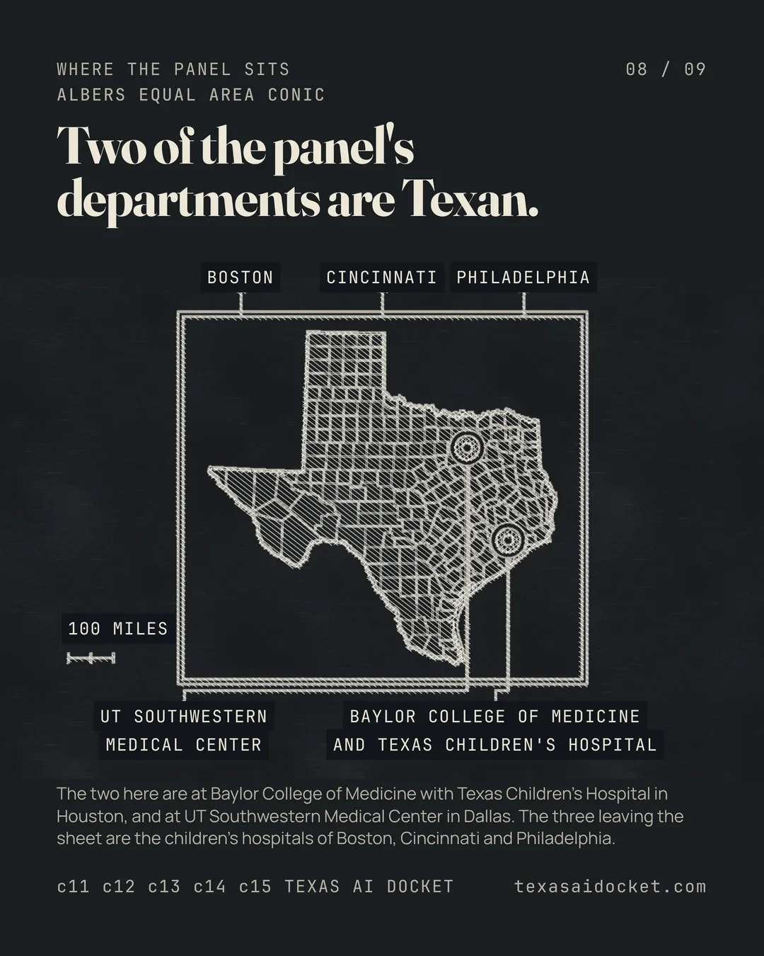
One of the panel's authors is in the radiology department shared by Baylor College of Medicine and Texas Children's Hospital in Houston.
Department of Radiology, Baylor College of Medicine and Texas Children's Hospital, Houston, Texas, USA.
Another is in the radiology department at UT Southwestern Medical Center in Dallas.
Department of Radiology, UT Southwestern Medical Center, Dallas, Texas, USA.
The panel also draws an author from the radiology department at The Children's Hospital of Philadelphia.
Department of Radiology, The Children's Hospital of Philadelphia (CHOP), Philadelphia, Pennsylvania, USA.
The panel also draws an author from the radiology department at Boston Children's Hospital.
Department of Radiology, Boston Children's Hospital, Harvard Medical School, Boston, MassachuseOs, USA.
The panel also draws an author from the radiology department at Cincinnati Children's Hospital Medical Center.
Department of Radiology, CincinnaD Children's Hospital Medical Center, University of CincinnaD College of Medicine, CincinnaD, Ohio, USA.
The second paper's title names it a machine learning analysis of MRI radiomics for survival prediction in brain metastases.
MRI Radiomics for Survival Prediction in Brain Metastases: A Machine Learning Analysis.
The second paper carries the same electronic article date as the first.
<ArticleDate DateType="Electronic"><Year>2026</Year><Month>09</Month><Day>09</Day></ArticleDate>
The second paper is registered under its own digital object identifier.
<ELocationID EIdType="doi" ValidYN="Y">10.3174/ajnr.A9630</ELocationID>
The paper frames the prognostic usefulness of radiomics in brain metastasis as still unclear.
Radiomics offers a means of extracting high-dimensional imaging biomarkers, but its prognostic utility in brain metastasis remains unclear.
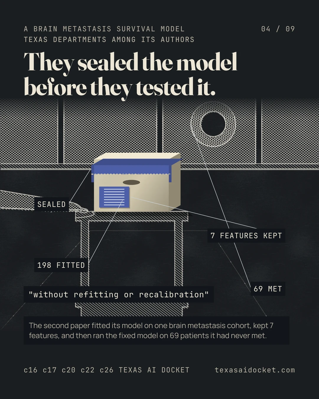
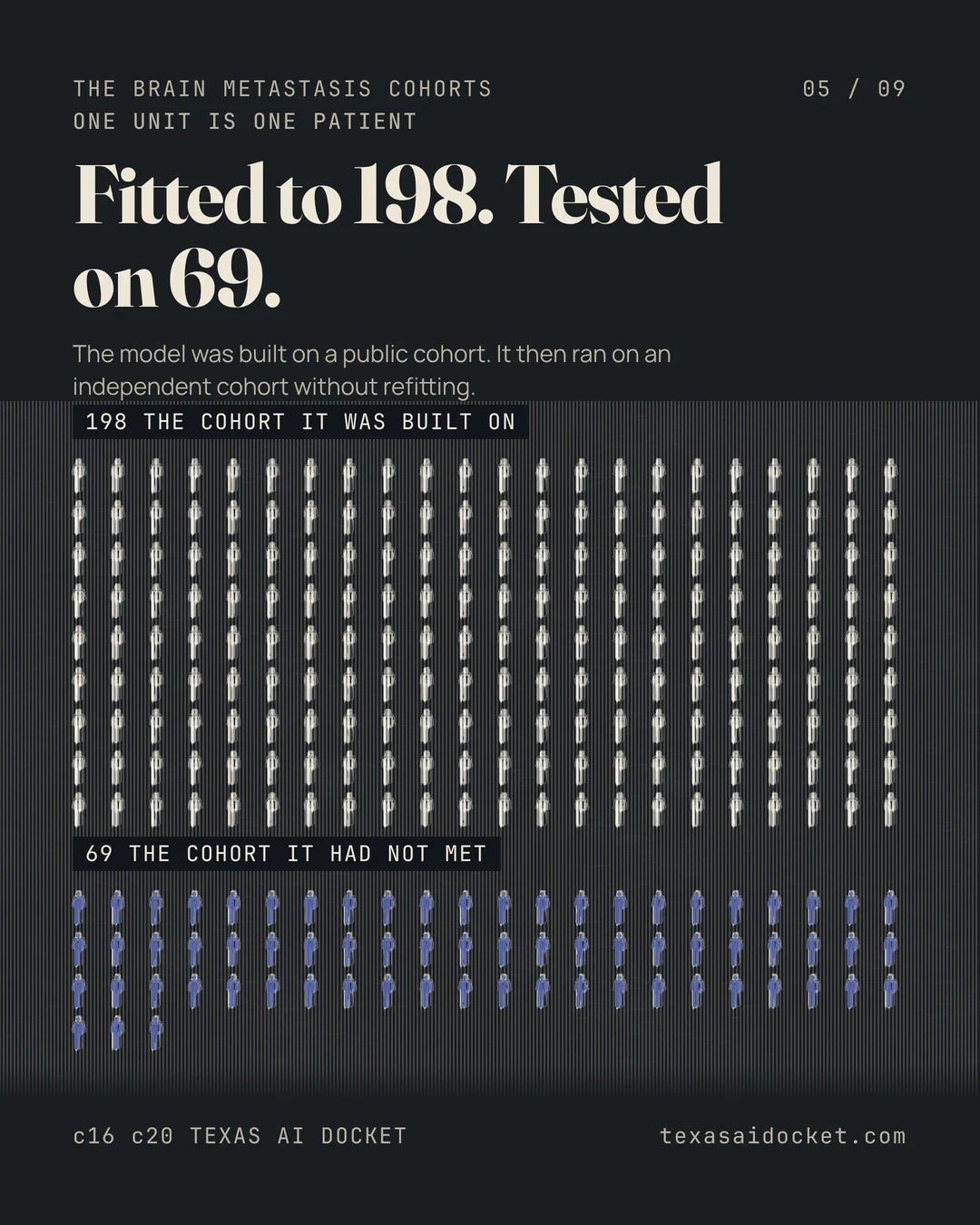
The model was built on a public cohort and then tested, without refitting, on an independent cohort.
This retrospective study developed a T1 postcontrast MRI radiomics survival model in a public brain metastasis cohort of 198 patients and externally validated the fixed radiomics model, without refitting or recalibration, in an independent cohort of 69 patients.
Discrimination was measured with the Harrell C-index and bootstrap resamples.
An elastic-net Cox model was selected with 10-fold cross-validation. Model discrimination was assessed with Harrell C-index and 2000 bootstrap resamples for 95% CIs.
The final model kept seven features, and its discrimination was lower on the outside cohort than on the cohort it was built on.
The final model retained 7 nonzero T1 postcontrast radiomics features after correlation filtering and elastic-net selection. The radiomics score had a C-index of 0.615 (95% CI, 0.543-0.687) in the training cohort and 0.574 (95% CI, 0.491-0.658) in external validation.
Adding the established clinical score did not lift the model back to its training figure.
In the external-validation subset with available Graded Prognostic Assessment, the combined radiomics plus Graded Prognostic Assessment model had a C-index of 0.591 (95% CI, 0.509-0.672).
The authors state the result as a limit rather than as a success.
T1 postcontrast MRI radiomics showed limited standalone discrimination for overall survival in patients with brain metastases.
The authors ask for cautious use and for models that carry more than imaging.
These results support cautious use of radiomics as an exploratory imaging biomarker and emphasize the need for integrated prognostic models that include clinical, treatment, molecular, and systemic disease variables.
Two of the paper's departments are in Texas. They are neuroradiology at MD Anderson in Houston and radiology at the Medical Branch in Galveston, and the author list carries departments in other states as well.
From the Department of Neuroradiology (R.E., H.A.Q., S.A., A.M., P.K.), The University of Texas MD Anderson Cancer Center, Houston, TX 77030, USA; Department of Radiology (E.C.), Duke University Medical Center, Durham, NC, USA; Department of Radiology (H.A.S., A.N., M.W.), The University of Texas Medical Branch, Galveston, TX, USA; Department of Radiology (J.D.R.), University of California, San Diego, San Diego, Calif; Division of Computational Pathology (S.B.), Department of Pathology and Laboratory Medicine, Indiana University School of Medicine, Indianapolis, IN, USA; Indiana University Melvin and Bren Simon Comprehensive Cancer Center, Indianapolis, IN, USA and Department of Computer Science (S.B.), Luddy School of Informatics, Computing, Engineering, Indiana University, Indianapolis, IN, USA; Medical Research Group, MLCommons, San Francisco, CA, USA.
The federal device regulator says the AI-enabled devices on its list met its premarket requirements, including a review of whether the studies suited the intended use.
The devices in this list have met the FDA's applicable premarket requirements, including a focused review of the device's overall safety and effectiveness, which includes an evaluation of study appropriateness for the device's intended use and technological characteristics.